
Ghatkopar, Mumbai
(+91) 9969026600
(+91) 9372887736
furiaeye@gmail.com
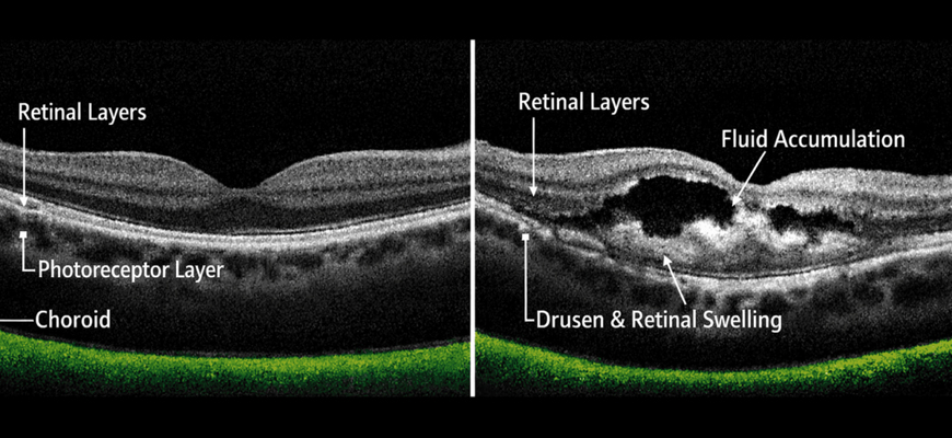
OCT (High Definition Retinal & Optic Nerve Scan)
Optical Coherence Tomography (OCT) is a non-invasive, high-resolution imaging technology that provides detailed cross-sectional scans of the retina and optic nerve. At FURIA'S EYE CLINIC, we use advanced OCT technology to detect, monitor, and manage a wide range of retinal and optic nerve conditions with exceptional precision.
OCT imaging allows our specialists to identify subtle changes in the eye that may not be visible through standard examination, enabling earlier diagnosis and more effective treatment planning.
What is OCT?
OCT uses light waves to capture detailed, layered images of the retina — the light-sensitive tissue lining the back of the eye — and the optic nerve. Similar to how ultrasound uses sound waves, OCT uses light to generate high-definition, cross-sectional views of eye structures in real time.
Conditions Diagnosed with OCT
- Age-related Macular Degeneration (AMD)
- Diabetic Macular Edema and Diabetic Retinopathy
- Glaucoma — detecting optic nerve damage and nerve fiber layer thinning
- Macular Hole and Macular Pucker
- Central Serous Retinopathy (CSR)
- Retinal Detachment
- Vitreoretinal Interface Disorders
- Optic Nerve Disorders
How is the OCT Scan Performed?
The OCT scan is a quick, painless, and non-contact procedure. The patient sits in front of the OCT machine and looks at a target light while the device scans the eye. The entire process takes only a few minutes and requires no injections or dye in most cases.
Steps Involved
- Pupil dilation may be performed for a wider field of view
- Patient is positioned comfortably in front of the OCT device
- The scan is performed — no contact with the eye
- High-resolution images are immediately available for analysis
Benefits of OCT Imaging
- Non-invasive and painless — no contact with the eye
- High-definition, detailed retinal and optic nerve imaging
- Early detection of subtle retinal and optic nerve changes
- Enables accurate monitoring of disease progression
- Guides treatment decisions for retinal and glaucoma conditions
- Quick procedure with immediate results
Why Choose FURIA'S EYE CLINIC?
- Advanced high-definition OCT technology
- Expert interpretation by experienced ophthalmologists
- Comprehensive analysis for retinal and optic nerve health
- Integrated with complete eye examination for accurate diagnosis
- Patient-friendly and efficient diagnostic process
Facilities Available

FURIA'S EYE CLINIC
FURIA'S EYE CLINIC is dedicated to providing comprehensive, compassionate, and advanced eye care for patients of all ages. With a commitment to excellence, our experienced ophthalmologists combine clinical expertise with modern diagnostic technology to deliver accurate diagnoses and effective treatments for a wide range of eye conditions.
call now
+91 9969026600
+91 9372887736
Facilities Available
- Stitchless Cataract Surgery (Phacoemulsification) & Lens Implantation
- Lasik Surgery for Refractive Error (Glasses)
- OCT (High Definition Retinal & Optic Nerve Scan)
- Topography & Pachymetry for Corneal Evaluation
- Retinal Laser, Injection & Surgery
- Pediatric Evaluation & Squint Surgery
- Cosmetic Eye Surgery
- Glaucoma Care & Surgery
- Perimetry
- Yag Laser
Contact Details
copyright © 2026 Furias Eye Clinic. all right reserved.